
 3 days Ago By Korea Bizwire
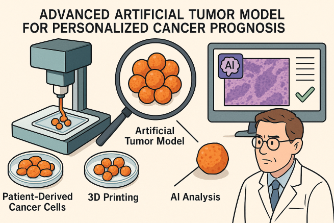
Breakthrough 3D-printed tumor tissue mimics real cancer environments, enabling AI to predict gene expression with 99% accuracy. (Image created by ChatGPT) ULSAN, April 30, (Korea Bizwire) — South Korean scientists have created an advanced artificial tumor model using 3D printing and artificial intelligence (AI) to replicate the internal environment of cancer patients’ bodies, offering a breakthrough in personalized cancer prognosis. The joint research team from the Ulsan National Institute of Science and Technology (UNIST) and Seoul Asan Medical Center announced Tuesday that they have developed an artificial cancer tissue, named Eba-PDO, that mimics the high-density, low-oxygen conditions characteristic of real tumors.
By analyzing the structure of Eba-PDO using AI, the team successfully predicted the expression of key prognostic genes for colorectal cancer with 99 percent accuracy. According to the researchers, cancer cells tend to grow in densely packed, oxygen-deficient environments, making tumors harder than normal tissues. Traditional patient-derived tumor models, however, have struggled to fully replicate these physical conditions, often leading to distorted results in studies of cancer cell growth and drug responses.

Patient-specific artificial cancer tissue bioprinting and AI-based patient prognosis prediction. (Image courtesy of Ulsan National Institute of Science and Technology.) To overcome these limitations, the team cultured patient-derived cancer cells three-dimensionally to form tumor organoids, mixed them with a specialized bio-ink made from gelatin and extracellular matrix components, and used 3D printing to create bead-like artificial tumors.
This method accurately recreated the stiffness and hypoxia seen in actual tumors. The genetic similarity between the artificial tumor tissue and real patient tumor samples reached 90 percent — a significant improvement over the conventional 70 percent. Moreover, the artificial model reliably reproduced individual variations in response to the chemotherapy drug 5-fluorouracil (5-FU).
Complementing this innovation, the team developed an AI model capable of predicting the expression of the CEACAM5 gene — a biomarker often found in colorectal and other solid tumors — solely from microscopic images. Overexpression of CEACAM5 is associated with increased metastasis and chemotherapy resistance. The AI was trained to recognize structural changes in the artificial tumor tissue, such as reduced cell adhesion and disrupted architecture, indicative of heightened CEACAM5 expression.
“Our approach, which reproduces real cancer cell growth outside the body, could lead to more precise, patient-tailored treatments,” a member of the research team said. “We expect to further enhance the model by integrating immune cells and vascular structures in the future.” The findings were published online on March 28, 2025, in the international journal Advanced Science .
The study was supported by Korea’s Ministry of Health and Welfare under the Korean ARPA-H initiative and by the Ministry of Trade, Industry and Energy’s Industrial Technology Alchemist Project. Ashley Song ( [email protected] ).